INFO/WWW-LINKS:
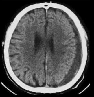
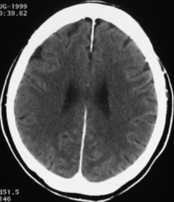
The
chronic haematoma is hypodense (= low attenuation) compared with the adjacent
brain.
Fresh
blood is high density compared with brain on CT and, at brain windows, appears
white. As the clot absorbs, the lesion becomes isodens (2-3 weeks after bleeding)
and, later, of low attenuation
Sites of intracranial heamorrhage/haematoma:
1) Between inner tabel and dura = extradural
heamorrhage/haematoma
2) Between dura and arachnoid = subdural
heamorrhage/haematoma
3) Between arachnoid and brain surface = subarachnoid
heamorrhage/haematoma
4) Within brain = intracerebral heamorrhage/haematoma
5) Within ventricles = intraventricular
heamorrhage/haematoma