INFO/WWW-LINKS:
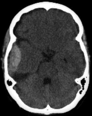
Fresh
blood is high density compared with brain on CT and, at brain windows, appears
white. As the clot absorbs, the lesion becomes isodens (2-3 weeks after bleeding)
and, later, of low attenuation
Sites of intracranial heamorrhage/haematoma:
1) Between inner tabel and dura = extradural
heamorrhage/haematoma
2) Between dura and arachnoid = subdural
heamorrhage/haematoma
3) Between arachnoid and brain surface = subarachnoid
heamorrhage/haematoma
4) Within brain = intracerebral heamorrhage/haematoma
5) Within ventricles = intraventricular
heamorrhage/haematoma