<<<
Compare
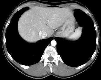
the pathological image-left and the physiological image-right
(blinded)
<<
F:
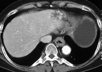
Stenosis
and wall-thickening in the distal third of the esophagus. Notize the 2 cm in
diameter well-circumscribed leasion of the left lobe of the liver with increased
contrast-enhancement from the border to the centre
H:
Adult
man, 51-years-old, with dysphagia, alcohol abuse
INFO/WWW-LINKS:
The onset of difficultiy in swallowing (progressing from solid to liquid bolus)
rises the suspicion of esophageal
cancer. Two-thirds of all patients with esophageal cancer are male. These
tumors are predominantly squamous cell carcinomas - except at the lower end
of the esophagus (adenocarcinoma).
A typical sign for hemangioma
of the liver is the delayed uptake of contrast-agent into the lesion, which
slowly fills from the border to the centre during minutes
D:
Esophageal
cancer
of the distal esophagus, hemangioma
of the left lobe of the liver
IN
THIS PART OF THE PAGE YOU FIND SOME TEXT FIELDS WHICH CAN BE OPENED EIGTHER
STEP BY STEP (CLICK ON "HISTORY", "HELP", "FINDINGS",
"DIAGNOSIS" OR "INFO/WWW-LINKS") OR AT ONCE WITH A CLICK
ON "ALL ON" - VICE VERSA CLICK ON "ALL OFF".
It is not
easy to find an exactly corresponding slice to every pathological example!
For that reason the
FILM
(2)
is
recommended!
Once opened you may use it for every pathological example.
If you need a physiological
image to compare click here